Electroencephalography (EEG): A Look at Understanding Brain Activity
In this article, we will examine in detail the basic operation and electrode structure of the EEG device as a reflection of the developments in the field of medical electronics. We will also focus on the importance of EEG in neurological research and the advantages of real-time monitoring of brain activity. We will explore how these technological advances are used to gain a deeper understanding of brain health.
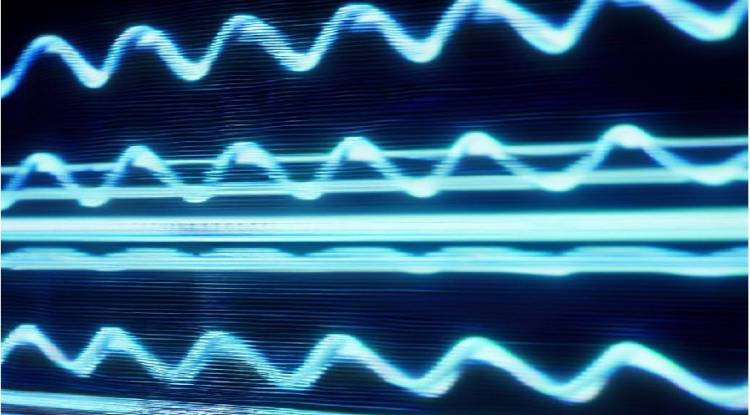
An electroencephalograph, or EEG, is a medical device used to measure and record brain activity. This device is used to capture and record the electrical activity produced by the brain. EEG is widely used, especially in areas such as epilepsy, neurological disorders, and sleep research.
The working principle of the EEG device is as follows:
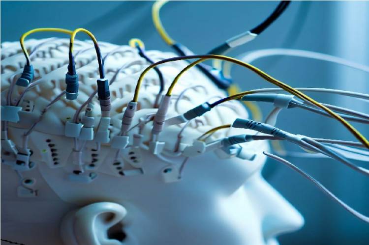
1. Electrodes: An EEG device contains small metal electrodes that are placed on the scalp of the patient or subject. These electrodes are used to detect weak electrical signals produced by the brain.
2. Amplification: These weak electrical signals from the electrodes are amplified by the amplifiers of the device. This amplification provides better visualization and analysis of signals.
3. Data Logging: After amplification, the device records the signals. This recording shows how brain activity changes over a period of time. The recorded data is usually transferred to a computer or a special recording device.
4. Data Analysis: The recorded data is then analyzed by experts. This analysis can help identify how brain activity changes or potentially abnormal activity over different periods. Abnormal electrical patterns can be detected, especially in cases such as a diagnosis of epilepsy.
EEG is an important medical tool used to monitor and evaluate brain activity in real time. Properly trained professionals can obtain valuable information about neurological disorders, seizures, sleep disorders, and other brain-related conditions by examining EEG data.
What is the structure of the electrodes?
EEG electrodes are electrodes made of thin metal tips used to measure and record brain activity. These electrodes are placed on the scalp and used to detect weak electrical signals from the brain. The structure of the electrodes is generally as follows:
1. Metal Tips: The most important part of the electrodes are the metal tips used to detect brain activity. These metal tips are usually made of silver or gold plated metals. These metals help to more precisely measure brain activity because of their high electrical conductivity.

2. Insulating Material: Metal leads are surrounded by an insulating material to ensure correct transmission of electrical signals. This insulating material is usually made of plastic or ceramic. The insulating material prevents different electrodes from touching each other and prevents unwanted signal distortions.
3. Connection Cables: The electrical signals from the electrodes are transmitted to the amplifiers of the device. Therefore, the electrodes are connected to the amplifier device with the appropriate connecting cable or wires. This wiring ensures reliable transmission of signals.
4. Application and Placement Mechanism: EEG electrodes should be placed on the scalp correctly. Therefore, there is an application and placement mechanism that keeps the electrodes in the correct position. This mechanism helps the electrodes to be firmly attached to the desired areas.
5. Gel or Paste: The electrodes can be coated with conductive materials such as gel or paste to provide better contact with the scalp and increase electrical conductivity. This is important to get more reliable and clear signals.
In general, EEG electrodes are special electrodes designed to measure brain activity. The design and material selection of these electrodes is carefully done to ensure reliable and precise measurements.
Brain waves : Brain Waves and EEG
Related Link: FDA Grants Neuralink Firm to Brain-Machine Interface Clinical Studies


